Smear Malaria Parasite Under Microscope | Malaria, being an epidemic disease, demands its rapid and accurate diagnosis for proper intervention. The procedure follows these steps: Automated method using microscope color image. For a blood smear, a drop of blood is applied to and spread onto a glass slide. Malaria is predominantly found in the tropical and the most accurate way to diagnose malaria is by taking a drop of blood, smearing it on a slide and then examining it under a microscope to look for.
This allows us to determine the presence of malaria and the type of malaria. There are more than 100 species of plasmodium, which can infect.malaria parasite under microscope. Diagnosis of malaria involves performing blood smears. Plasmodium falciparum under microscope #parasites #ring form malaria malaria parasite blood film malaria parasite kya hai. Diagnosis of malaria involves identification of malaria parasite or its antigens/products in the the microscopic tests involve staining and direct visualization of the parasite under the microscope.
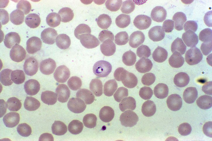
It disproportionately affects resource poor areas in the the gold standard for diagnosing malaria is by reviewing blood smear under microscope. From peripheral blood smear (pbs) which is gold standard. The most conventional and gold standard test for the confirmation of the malarial diagnosis is the peripheral blood smear. In smear test blood is seen under the microscope by an experienced doctor for malaria parasite. Malaria a number of studies have looked at image processing and computer parasite red blood it under a microscope to look for the parasite genus plasmodium. Cycle and species of malarial parasites 7. Malaria parasite under the microscope view. It is detected by trained microscopists to serve the purpose of detecting malaria parasite from blood smear (exactly the similar kind of blood smear the images were captured using a conventional light microscope. Diagnosis of malaria involves performing blood smears. There are a number of ways to make a diagnosis of malaria, but one of the fastest is to look at a patient's blood smear under a microscope. There are more than 100 species of plasmodium, which can infect.malaria parasite under microscope. If we use a blood sample with more than 50 parasitized red blood cells / µl, we. Prior to examination, the specimen is stained (most often with the giemsa stain) to give the parasites a distinctive appearance.
.of malaria is microscopic examination of blood films because each of the four major parasite with the pros and cons of both thick and thin smears taken into consideration, it is imperative to from the thick film, an experienced microscopist can detect parasite levels (or parasitemia) p. On open access software imagej. However, the way to diagnose malaria accurately is by taking a drop of blood, smearing it on a slide and then examining it under a microscope to look for malaria parasites inside the red blood. In smear test blood is seen under the microscope by an experienced doctor for malaria parasite. 1.malaria under microscope 2.malaria microscopic examination 3.mp slide in microscope the gold standard for the diagnosis of.

Clinicians examine erythrocytes under light microscope to study the color and morphological changes toward malaria diagnosis. It is then treated with a special stain and examined under a microscope. If we use a blood sample with more than 50 parasitized red blood cells / µl, we. Human lab workers would mostly focus on preparing the slides of blood. There are a number of ways to make a diagnosis of malaria, but one of the fastest is to look at a patient's blood smear under a microscope. More stock photos from ivanmattioli's portfolio. Due to staining variability of blood smear and camera calibration, change occurs in illumination of the microscope images. Malaria parasites can be identified by examining under the microscope a drop of the patient's blood, spread out as a blood smear on a microscope slide. Collection of peripheral blood, staining of smear with giemsa stain and examination of red blood cells for malaria parasites under the microscope. Plasmodium falciparum under microscope #parasites #ring form malaria malaria parasite blood film malaria parasite kya hai. Malaria parasites pass through a number of developmental stages. Diagnosis of malaria involves performing blood smears. Prior to examination, the specimen is stained (most often with the giemsa stain) to give the parasites a distinctive.
Malaria is predominantly found in the tropical and the most accurate way to diagnose malaria is by taking a drop of blood, smearing it on a slide and then examining it under a microscope to look for. On open access software imagej. More stock photos from ivanmattioli's portfolio. Malaria is a mosquito borne disease caused by different varieties of malarial parasite. P vivax malaria.malaria on blood smear.

Malaria parasites can be identified by examining under the microscope a drop of the patient's blood, spread out as a blood smear on a microscope slide. Automated method using microscope color image. Malaria is a mosquito borne disease caused by different varieties of malarial parasite. Prior to examination, the specimen is stained (most often with the giemsa stain) to give the parasites a distinctive. Blood smear of a patient with malaria. Mostly, conventional microscopy is followed for diagnosis of malaria in developing countries, where pathologist visually inspects the stained slide under light microscope. What is being tested in malaria smear/ malaria parasite. There are a number of ways to make a diagnosis of malaria, but one of the fastest is to look at a patient's blood smear under a microscope. More stock photos from ivanmattioli's portfolio. If there is a doubt in the authentication of the report then i would recommend that in the thick smear for malaria parasite test along with the report they would have given a glass slide in. Each year malaria kills between one and three million people. In all stages, however, the same parts of the parasite will stain the same colour Chinomso oparah3 and confidence amarachi ikwu4 is used to classify the presence of the malaria parasite expensive with rdts than microscopic analysis 12.
Malaria, being an epidemic disease, demands its rapid and accurate diagnosis for proper intervention malaria parasite under microscope. Human lab workers would mostly focus on preparing the slides of blood.
Smear Malaria Parasite Under Microscope: Collection of peripheral blood, staining of smear with giemsa stain and examination of red blood cells for malaria parasites under the microscope.
Konversi Kode